Advertisements
Magnetic Resonance Imaging (MRI) has undeniably transformed the landscape of modern healthcare. It’s a non-invasive technology that provides remarkable insights into the body’s internal workings, all without relying on harmful radiation. In a world where precision is paramount, MRI offers detailed, three-dimensional images of soft tissues, organs, and even the brain. This technology is indispensable for diagnosing and monitoring a wide range of conditions—from brain injuries to cancers, and beyond.
At its core, MRI works by utilizing strong magnetic fields and radio waves. Unlike X-rays or CT scans, MRI doesn’t rely on ionizing radiation, which is a major advantage in terms of patient safety. Instead, MRI uses a magnetic field to align protons in the body’s tissues. A pulse of radio waves temporarily knocks these protons out of alignment, and as they realign, they emit energy. This energy is captured by sensors in the MRI machine and converted into highly detailed images. It’s a fascinating and elegant process that allows healthcare professionals to visualize what’s happening deep within the body.
Advertisements

One of the standout features of MRI is its ability to capture incredibly detailed images of soft tissues. While X-rays and CT scans are excellent for viewing bones, MRI shines when it comes to muscles, ligaments, and the brain. This is why MRI is so frequently used to assess conditions that affect the brain and spinal cord, such as tumors, injuries, and degenerative diseases. It’s also incredibly valuable for imaging the heart, liver, and other organs, giving doctors a clear view of how they’re functioning.
What makes MRI even more powerful is its versatility. There’s functional MRI (fMRI), for instance, which tracks brain activity in real-time. fMRI has revolutionized the way we understand brain function, allowing scientists and doctors to see how the brain responds to different tasks. Whether it’s a patient undergoing surgery or a researcher studying cognitive processes, fMRI provides invaluable insights into the brain’s inner workings.
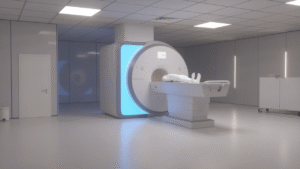
The MRI procedure itself is quite simple: the patient lies down on a table, which slides into a large, tube-like machine. For many, this can be a daunting experience, particularly due to the confined space and the loud noises the machine makes during the scan. However, newer machines, such as open MRIs, aim to alleviate these discomforts by providing a less claustrophobic experience. Despite these advances, some patients still require relaxation techniques or even sedation for more extended scans.
Safety is one of MRI’s most appealing aspects. Since it doesn’t use ionizing radiation, it’s considered much safer than X-rays and CT scans. There’s no risk of radiation exposure, which makes MRI an excellent option for monitoring ongoing conditions or performing routine check-ups. However, the powerful magnetic field means that patients with metal implants—such as pacemakers or prosthetic joints—must disclose this information before undergoing the procedure. For those with metal in their body, MRI might not be an option, or it may require special precautions.
On the flip side, there are some limitations. One of the primary concerns is cost—MRI machines are expensive to acquire and maintain, which can make them less accessible, especially in low-resource settings. Additionally, while MRI is widely available, not every facility has the advanced machines necessary to perform certain types of scans. This discrepancy can lead to delays in diagnosis, which can be critical in urgent cases.
There’s also the noise. The sounds produced by an MRI machine can be loud and unsettling. It’s not unusual for patients to feel anxious during the procedure. Thankfully, patients are usually given earplugs or headphones to help block out the noise. For those particularly sensitive to sound, the experience can still be uncomfortable, but it’s a small price to pay for the level of detail MRI provides.
Looking ahead, the future of MRI is incredibly exciting. With the integration of Artificial Intelligence (AI), MRI technology is poised to become even more advanced. AI is being used to analyze MRI scans faster and more accurately, reducing human error and helping doctors make quicker, more informed decisions. Machine learning algorithms are already showing promise in detecting subtle abnormalities that may be overlooked by the human eye.
Additionally, research into new contrast agents and improved imaging techniques continues to push the boundaries of what MRI can do. In particular, advancements in magnetic resonance elastography (MRE) are helping doctors assess tissue stiffness, offering a more detailed view of organs like the liver without the need for invasive biopsies. This could open the door for non-invasive, early detection of conditions like liver disease and cancer.
MRI is a remarkable tool, and as technology continues to evolve, it promises even more breakthroughs in medical diagnostics. It’s not just about seeing inside the body—it’s about understanding it in ways that were previously unimaginable. For patients, MRI offers hope, precision, and peace of mind. And for the medical community, it’s a technology that has forever changed the way we approach healthcare.
Related MRI Professional Information
| Name | Dr. John Doe |
|---|---|
| Profession | MRI Specialist, Radiologist |
| Experience | 15+ years in MRI diagnostics |
| Academic Background | MD, Radiology Residency, University of Harvard |
| Current Role | Senior MRI Consultant at XYZ Medical Center |
| Research Focus | Advanced MRI imaging techniques and AI integration in diagnostics |
| Website | MRI National Institute |
For more detailed information on MRI scans and advancements in the field, visit the National Institute of Biomedical Imaging and Bioengineering.
Advertisements